Presentation
Knee deformity after trauma.
Patient Data
Age: 14 years
Gender: Female
From the case:
Alar ligament of the patella injury


Download
Info
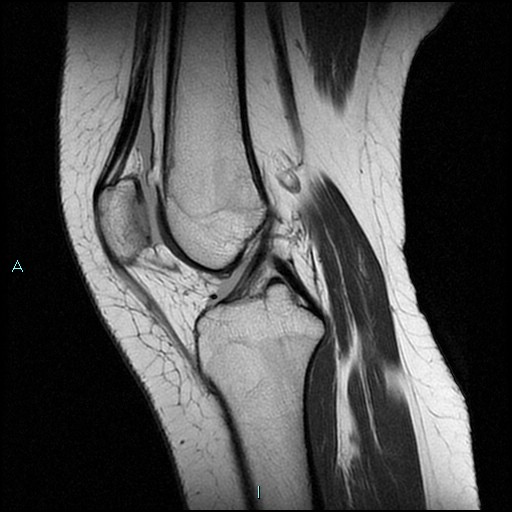
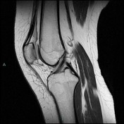
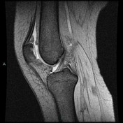
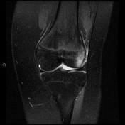
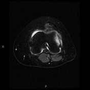
MRI knee
Medial alar ligament lesion with external patellar subluxation. Bone marrow edema is present in the medial surface of the patella and in the lateral femoral condyle. The femoral trochlea is flat. There is a knee joint effusion.
Normal cruciate ligaments, collateral ligaments, and menisci.
Case Discussion
The patella is maintained in place by two ligaments one internal and external and are called the alar ligaments.
After the first episode of dislocation, the possibility of recurrence is high. This is especially the case if dislocation occurs at a young age or if there are predisposing congenital factors, such as a valgus knee, or flattened trochlea.


 Unable to process the form. Check for errors and try again.