
Presentation
Chronic left nasal obstruction and discharge
Patient Data
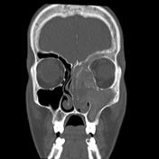

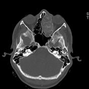
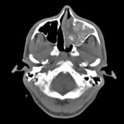
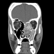
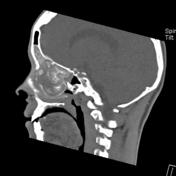

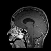
Expanded left maxillary antrum, left ethmoidal complexes and the left side of the frontal sinus. They show complete inhomogeneous opacification with hyperdense material is seen located centrally surrounded by hypodense mucosa.
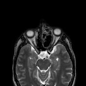
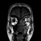
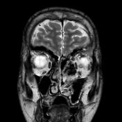
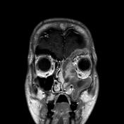

The examined sinuses show internal high signal at T1 WI and signal void at T2 WI.
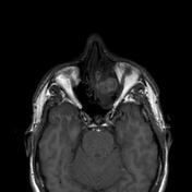
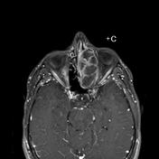
Contrast enhancement of the mucosal lining.
No intra-cranial or intra-orbital extension.
Case Discussion
Allergic fungal sinusitis (AFS) is the most common form of fungal sinusitis and is common in warm, humid climates.
The majority sinuses show near complete opacification. On unenhanced CT the sinuses are typically opacified by centrally (often serpentinous) hyperdense material with a peripheral rim of hypodense mucosa.
Approximately 40% of patients may have each of the following features:
- expansion of an involved sinus
- remodeling and thinning of the bony sinus walls
- erosion of the sinus wall.


 Unable to process the form. Check for errors and try again.