
Presentation
Progressive impairment of recent/short-term memory with relative preservation of remote and autobiographical memory.
Patient Data
Note: This case has been tagged as "legacy" as it no longer meets image preparation and/or other case publication guidelines.


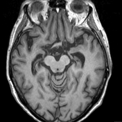
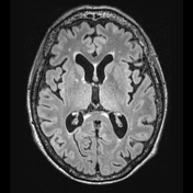
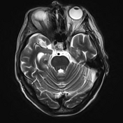
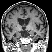

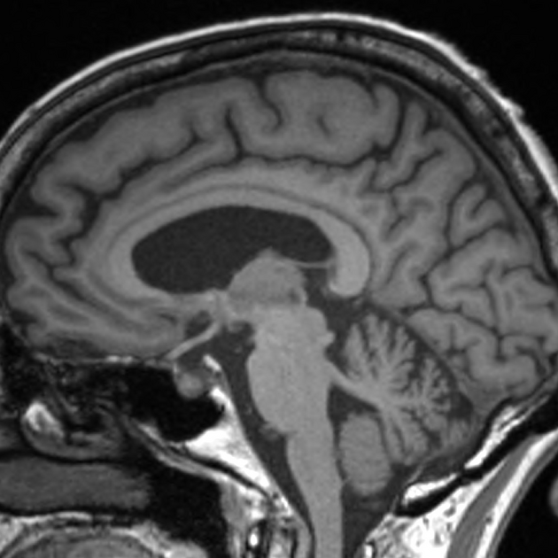
The sulci and ventricles are mildly more prominent than expected for age. The mesiotemporal structures, including the temporal horns of the lateral ventricles and the collateral sulci, are slightly more prominent, with bilateral hippocampus, amygdala and entorhinal cortex reduced in volume.
Multiple FLAIR hyperintense lesions in the supratentorial white matter, confluent in the periventricular regions, are in keeping with mild to moderate small vessel ischemia.
No diffusion restriction or suspicious susceptibility.
Case Discussion
Findings are typical of the clinically suspected mild Alzheimer disease or limbic-predominant age-related TDP-43 encephalopathy (LATE).
Unfortunately, these two entities can not only mimic each other clinically and radiologically but can also co-exist. An amyloid PET can be useful but currently, a definite antemortem diagnosis cannot be reached.


 Unable to process the form. Check for errors and try again.