
Presentation
Back pain
Patient Data
Age: 35 years
Gender: Male
From the case:
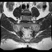
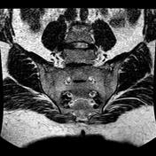
Ankylosing spondylitis "Romanus lesions"


Download
Info

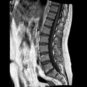
The examined lumbar vertebrae show small subchondral fatty marrow degenerative changes of the anterior aspects of their upper and lower endplates "Romanus lesions", they elicit high signal at T1 and T2 WI giving the "Shiny corner sign"
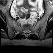
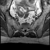
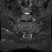
Severe narrowing mounting to near total ankylosis of the anterior aspects of both sacroiliac joints with patchy areas of altered marrow signal.


 Unable to process the form. Check for errors and try again.