
Anterior cruciate ligament mucoid degeneration and ganglion cyst
Presentation
Pain and stiffness for 4 months. Old history of injury.
Patient Data
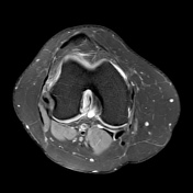

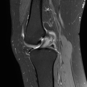

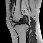
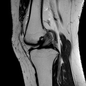

Thickened anterior cruciate ligament with celery stalk appearance and T2/PD fat sat intrasubstance hyperintense signals reflecting mucoid degeneration of the anterior cruciate ligament.
A T2 hyperintense cyst of 19 x 18 mm in the intercondylar fossa adjacent to the femoral attachment of anterior cruciate ligament, consistent with ganglion cyst.
A horizontal tear is noted at the anterior horn of the lateral meniscus.
Case Discussion
The etiology of ACL ganglion cysts is uncertain. Remote ligamentous trauma and mucoid degeneration play a role in the development of the cysts. MR imaging appearances are discrete fluid signal cystic structure within or on the surface of ACL. They have a tendency to extend into surrounding intra-articular or osseous structures.
Diffuse thickening with intra-ligamentous T2 hyperintensity and splaying of intact ACL fibers with a celery stalk appearance is consistent with mucoid degeneration of anterior cruciate ligament.


 Unable to process the form. Check for errors and try again.