
Arachnoid cyst
Updates to Study Attributes
no intra
axial-axial or extraaxial hemorrhage.-axial haemorrhageno calvarial fracture
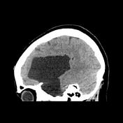
.large extra
axial-axial CSF attenuating lesion is seen occupying the left middle cranial fossa splaying thesylvianSylvian fissure and displacing the frontal, parietal and temporal lobes.secondary mass effect is seen in the form of effacement of left cerebral hemispheric sulci, mild midline shift to right,
effacedeffaced ipsilateral lateral ventricle.no transtentorial herniation
.
Image CT (non-contrast) ( update )
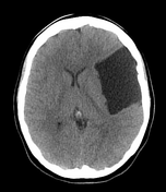
Image CT (non-contrast) ( update )
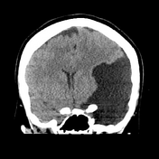
Image CT (non-contrast) ( update )
Updates to Study Attributes
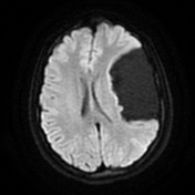
Contrast enhanced-enhanced MRI confirms
non
enhancing-enhancing nature of the lesion.CSF attenuation
.-
extra
axial-axial locationdisplaced cortical mantle and cortical vessels between the lesion and calvarium
no underlying white matter changes in the left cerebral hemisphere
.
Image MRI (DWI) ( update )
Updates to Case Attributes
Most common site of origin of the arachnoid cyst is in the middle cranial fossa, splaying the Sylvian fissure. Depending on the size and extent, it is classified by the Galassi classification. This is a type III variant.
They tend to be asymptomatic and pounced upon incidentally. The larger ones may cause symptoms secondary to mass effect.


 Unable to process the form. Check for errors and try again.