
Presentation
Lower limb weakness
Patient Data

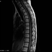
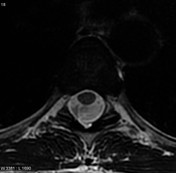

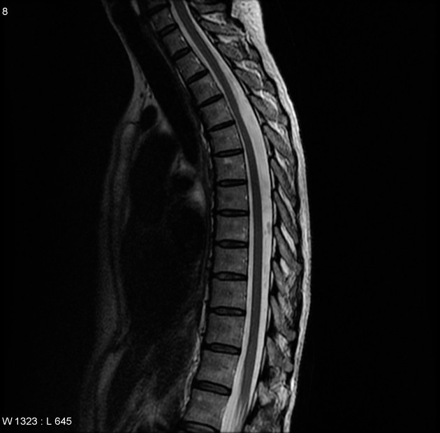
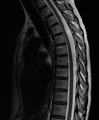
MRI of the spine demonstrates the upper thoracic cord to be displaced anteriorly, focally abutting the posterior aspect of the vertebral column. Axial T2 images suggest that the cord is being pushed anteriorly by a CSF intensity mass, rather than pulled forward.
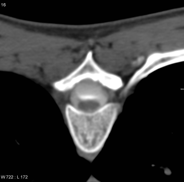
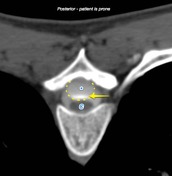
The patient went on to have a myelogram, which demonstrates contrast pooling in the dependent portions of a cystic structure with imperceptible walls, located posterior to the thoracic cord.
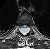
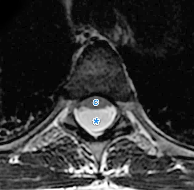
The thoracic spinal cord (C) is displaced anteriorly by a CSF intensity space ( * ) which on axial imaging in fact has slightly higher and more homogeneous signal than one normal sees in thoracic CSF.
Myelography, obtained with the patient prone, outlines the arachnoid cyst (yellow dotted line) with hyperdense contrast seen pooling dependently (yellow arrow).
Case Discussion
This case illustrates the typical appearances of a spinal arachnoid cyst, and also demonstrates how challenging it can sometimes be to confirm the diagnosis.


 Unable to process the form. Check for errors and try again.