
Presentation
Adult male with a long history of dyspnea and exercise intolerance. ECG revealed a bundle branch block
Patient Data


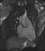
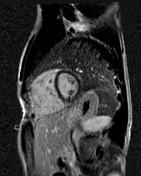

Multiple cine sequences show an enlarged right ventricle, abnormal right ventricle wall motion including dyskinesia and paradoxical movement most notable at the segment connecting the RVOT to the right ventricular apex. There is also abnormal corrugation of the right ventricle apex. No abnormal enhancement on the late gadolinium sequence. The 4 CH cine is compromised by the patients' arrhythmia.
Case Discussion
Arrhythmogenic right ventricular cardiomyopathy (ARVC) is characterized by progressive replacement of normal myocardium in the right ventricle by fibrofatty tissue, this finding can be depicted on MRI, however, the histopathology is the most reliable tool. The most common location for the fibrofatty changes are the RVOT, right ventricle apex and the inferior surface of the right ventricle 1.
The diagnosis of ARVC is based on the presence of major and minor criteria according to the revised Task Force Criteria 2010. The diagnostic criteria include MRI, echocardiography, ECG, and histology.
The rule of MRI is to study the structure and function of the RV. In our case there are two major criteria include decreased RVEF < 40 %, and increased RV index ( EDV/ BSA ) is 116. In addition to abnormal right ventricular wall motion, in the form of paradoxical movement of the segment connecting the RVOT to the right apex.
Revised Task Force MRI Criteria 2010
MAJOR Criteria
- Regional RV akinesia or dyskinesia or dyssynchronous RV contraction
- AND 1 of the following
RVEDV index > 110 mL/m2 (male)
RVEDV index > 100 mL/m2 (female)
RVEF < 40%
MINOR Criteria
- Regional RV akinesia or dyskinesia or dyssynchronous RV contraction
- AND ONE of the following
RVEDV index > 100 to <110 mL/m2 (male)
RVEDV index > 90 to < 100 mL/m2 (female)
RVEF > 40% to < 45%
Definite: 2 Major, or 1 Major + 2 Minor, or 4 Minor
Borderline : 1 Major + 1 Minor, or 3 Minor
Possible : Major or 2 Minor


 Unable to process the form. Check for errors and try again.