
From the case:
Arytenoid cartilages (illustration)

Download
Info

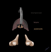
Arytenoids illustrated in context of other structures nearby.
Author: Candace Makeda Moore, MD
License: CC-NC-BY-SA
Case Discussion
Arytenoid cartiliges are illustrated in the first image (in white) as well as thyroid , cricoid and corniculate cartilages. In this image the cartilages are shown in anatomical position at top, then shows as rotated forward atop the cricoid. In the second image a view as if in the middle of an intubation is drawn with a laryngoscope for orientation.


 Unable to process the form. Check for errors and try again.