
Presentation
Pain and swelling of the neck.
Patient Data
Age: 25 years
Gender: Male
From the case:
Atypical 2nd branchial cleft cyst (type IV) - infected


Download
Info
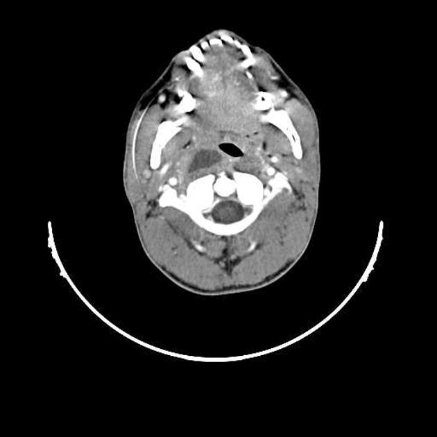
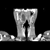
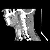
Well-defined cystic mass with thick, smooth and enhanced wall is seen at parapharyngeal-prevertebral spaces. Cervical lymphadenopathies also are noted.
Case Discussion
The imaging findings are compatible with 2nd branchial cleft cyst (atypical - type IV) which seems to be infected. Although the most common location for a 2nd branchial cleft cyst is the submandibular space, the cyst can atypically present in the parapharyngeal space.


 Unable to process the form. Check for errors and try again.