
Presentation
Hypo- and hyperechoic left renal lesions on ultrasound.
Patient Data


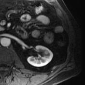
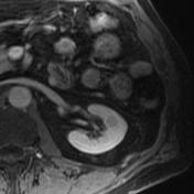
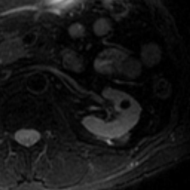
T1-hyperintense and T2-signal void cyst suggestive of hemorrhagic or proteinaceous content. No enhancement after administration of IV gadolinium.
Case Discussion
There is a small cyst in the left kidney that is T1 hyperintense and not enhancing, compatible with a hemorrhagic cyst.
There is also a multilobulated cyst which shows no measurable enhancement of its septa and no mural nodularity. Evaluation for calcification is limited on MRI, but there is no obvious calcification.
The appearance is compatible with minimally complicated cysts, without worrisome features. This would be analogous to a Bosniak II cyst, but strictly speaking, the Bosniak criteria are meant to be used with CT imaging.


 Unable to process the form. Check for errors and try again.