
Presentation
Dyspareunia
Patient Data
Age: 45 years
Gender: Female
From the case:
Bartholin gland cyst


Download
Info

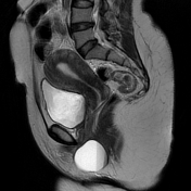
There is a well-defined unilocular cystic lesion arising from the left lateral wall of the lower vagina at and below the pubic symphysis. It appears of low signal intensity on T1WI, high signal intensity on T2WI with hypointense fluid-fluid levels (chronic blood products). No peripheral enhancement seen on postcontrast sequences.
Uterus and both ovaries show normal appearance.
Case Discussion
MRI features of a Bartholin gland cyst.
On imaging, the differential diagnosis includes:
- Bartholin gland abscess: usually there is associated inflammatory features
- Bartholin gland tumor: usually seen in the post-menopausal patient
- Gartner duct cyst: located at or above the level of the pubic symphysis
- Nabothian cyst: located within the uterine cervix
- Skene duct cyst: closer to the external urethral meatus
- urethral diverticulum: usually the patient presents lower urinary tract symptoms


 Unable to process the form. Check for errors and try again.