
Presentation
Left loin pain associated with nausea and vomiting.
Patient Data
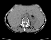

The elongated left lobe of the liver extends across the midline into the left hypochondrium lying in close contact with the spleen (beaver tail liver--anatomical variant).
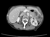
A 16 mm well-defined lesion containing soft tissue and fat densities, likely an angiomyolipoma along the posterior aspect of the upper/mid pole of left kidney is visualized.
Bilateral prominent extrarenal pelvis and retro-aortic left renal vein (type 1) noted.
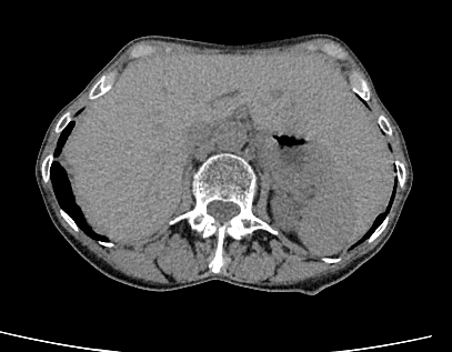
Mild diffuse mural thickening of the urinary bladder along with few small bladder diverticula seen.
Case Discussion
Beaver tail liver is a normal variant of hepatic morphology, which is more common in the females. Prominent extrarenal pelvis and retroaortic left renal vein are also anatomical variants.


 Unable to process the form. Check for errors and try again.