
Presentation
Chronic headache
Patient Data
Age: 50 years
Gender: Female
From the case:
Benign notochordal cell tumor


Download
Info
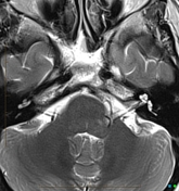
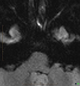
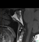
Elongated band-like lesion in central clivus, dark on T1, bright on T2.
From the case:
Benign notochordal cell tumor
Download
Info
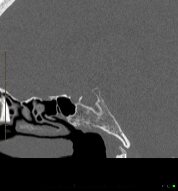
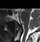
CT is almost normal, with subtle sclerosis in the region of MR abnormality.
From the case:
Benign notochordal cell tumor
Download
Info
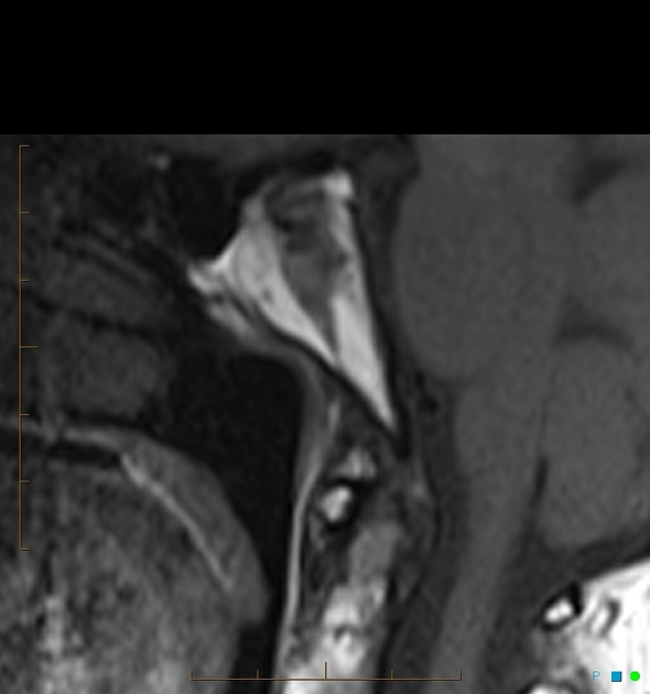
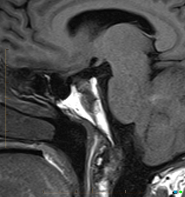
MR cervical spine from 5 years prior with Sagittal T2 to better illustrate the bright T2 signal. Lesion has been stable.
Case Discussion
Not many differential diagnosis is available. Benign persistent notochordal tissue is one of the possibilities. Various evolving terms have been used in the literature e.g. notochordal rests, tumor, hamartoma - all suggest benignity.


 Unable to process the form. Check for errors and try again.