
Presentation
Intermittent lower abdominal pain since 2 months. Irregular menstrual cycles.
Patient Data
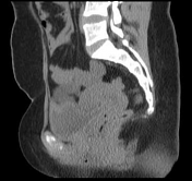

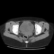

Two discrete uterine horns identified. Endometrium and cervical canal cannot be commented upon.
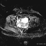
Left ovarian cyst is appearing hypo-attenuating.
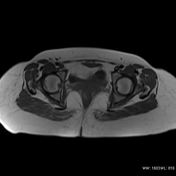

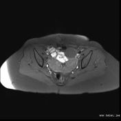
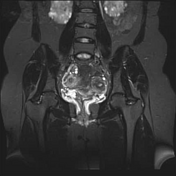
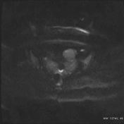

Two discrete uterine horns are noted. Fundal cleft measures more than 1 cm with widened intercornual distance. Separate non-communicating endometrial cavities seen.
Also seen is incomplete septum within the cervix. The vagina appears normal.
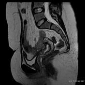
Left ovary shows a thin-walled cystic lesion with minimal internal debris and incomplete septae within. No evidence of any obvious mural nodule / solid component.
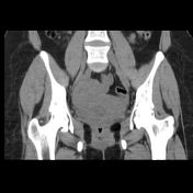
No suspicious lymphadenopathy.
Incidental bilateral polycystic kidneys noted.
Case Discussion
Bicornuate uterus with incomplete septum within the cervix.
Special thanks: Dr. Nanda Kumar


 Unable to process the form. Check for errors and try again.