
Presentation
A female presented with infertility.
Patient Data


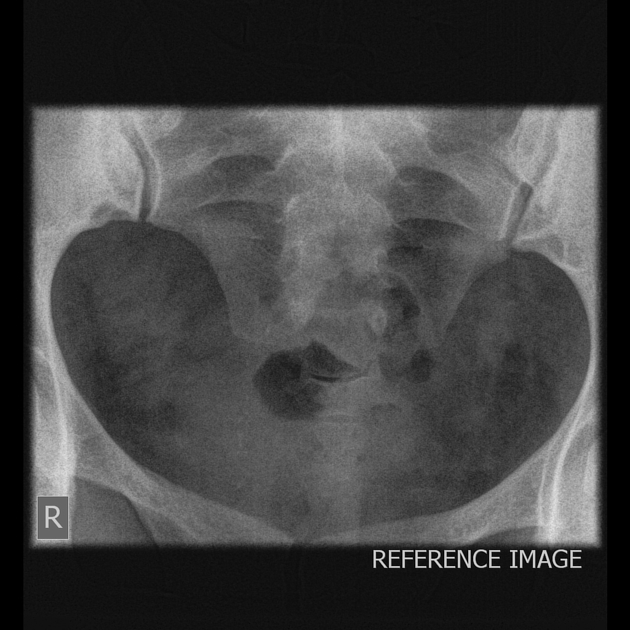
First image shows the reference or scout image.
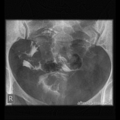
Second image shows opacification of two diverging endometrial cavities with two separate cervices with spillage of contrast into the peritoneal cavity through both horns.
Third image shows evidence of retained contrast within peritoneal cavity in delay image (25 minutes).
Case Discussion
Any disruption of Müllerian duct development can result in a broad spectrum of congenital abnormalities called Müllerian duct anomalies (MDAs).
Diagnosis of MDAs is clinically important because of the high associated risk of infertility, endometriosis, and miscarriage. MDAs are also commonly associated with renal anomalies.
Hysterosalpingography (HSG) is used in an initial evaluation of infertility; it allows assessment of the uterine cavity and fallopian tube patency but is deficient in providing any information about the external uterine contour. Currently, however, MR imaging remains the preferred MDA imaging method, as it exquisitely details both the uterine cavity and external contours and has shown excellent agreement with clinical MDA subtype diagnosis


 Unable to process the form. Check for errors and try again.