
Bilateral cavernous segments internal carotid artery aneurysms
Presentation
Acute onset of severe headache and blurred vision with background history of hypertension.
Patient Data
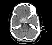

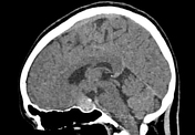

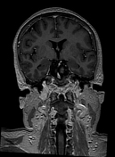
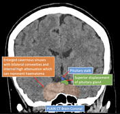
Abnormal high attenuating content within the right cavernous sinus as well as convexities in lateral walls of both cavernous sinuses. The sella turcica is widened and expanded.
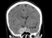
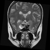
On coronal view, the pituitary gland and pituitary infundibulum appear to be displaced superiorly by the "mass/collection" within the sella turcica or cavernous sinus.
Optic chiasm is compressed.
From the non-contrast CT brain study, the identification of sella turcica/cavernous sinuses mass with possible hemorrhagic content raise suspicion of possible two differential diagnoses, either hemorrhagic pituitary macroadenoma with lateral cavernous sinuses extension or internal carotid arteries (ICA) aneurysm.
Proceeded with MRI brain/pituitary gland.

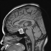
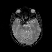
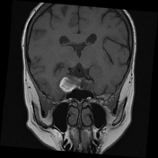
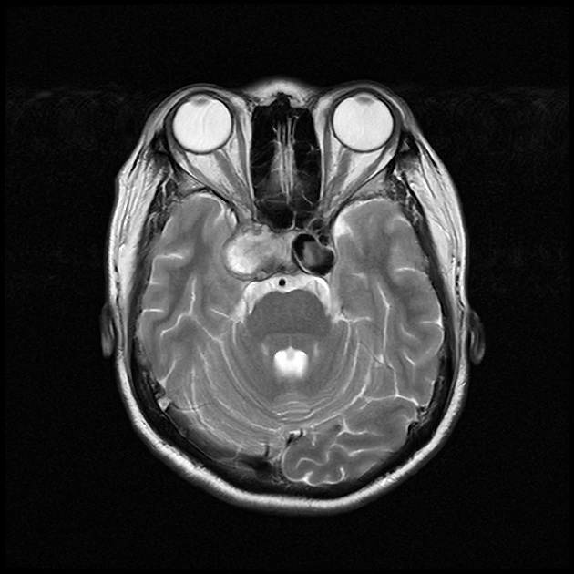
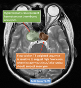
A well circumscribed spherical shaped mass with abnormal internal and peripheral flow voids occupying the left cavernous sinus. It appears to be in continuity with the cavernous segment of left internal carotid artery as saccular aneurysm. This mass enhances avidly as artery in post contrast study.
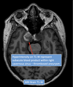
Abnormal hyperintensity on T1-weighted sequence seen within the right cavernous sinus (larger on the right side), left cavernous sinus as well as intercavernous sinus, suggestive of blood product. This is further confirmed by abnormal blooming artefacts on gradient echo sequence within the cavernous sinus.
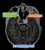
The T1-hyperintense "mass"/"collection" with epicenter at right cavernous sinus is expansile and causing significant mass effect as depicted by convexity on the right lateral wall of cavernous sinus and superior compression onto the optic chiasm. Absence of contrast opacification and flow void within the right cavernous "mass" may represent thrombosed right cavernous segment ICA aneurysm.
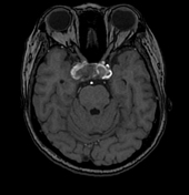
The mass effect of the thrombosed cavernous segment ICA within the right cavernous sinus suggests compression of the cranial nerves within it, namely, right oculomotor nerve (III), right trochlear nerve (IV), V1 and V2 of right trigeminal nerve (V) as well as the medially located right abducens nerve (VI). Posteriorly, no extension into the right Meckel's cave. Intracranial optic nerves are displaced superiorly by the aneurysm/hematoma.
Normal pituitary gland and posterior pituitary bright spot are not well observed, likely due to significant compression by the thrombosed aneurysm/hematoma.
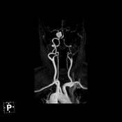
MRA of circle of Willis and carotid arteries:
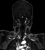
The major branches from aortic arch are right brachiocephalic artery, left common carotid artery and left subclavian artery. Both vertebral arteries arise from respective subclavian arteries.
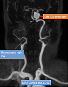
Minimal opacification of the C1 (cervical) segment of right internal carotid artery. The rest of segments (C2 till C6) of right ICA are not opacified, suggestive of thrombosis.
The C7 (communicating) segment of right ICA is well opacified with blood supply from the left anterior cerebral artery via the anterior communicating artery.
Bilateral common carotid arteries, right external carotid artery, left internal carotid artery and left external carotid artery are well opacified and normal in caliber.
Right ACA and right MCA are smaller in caliber. Normal caliber and opacification of left ACA, left MCA and anterior communicating artery.
Dominant left vertebral artery. Posterior vertebrobasilar circulation appears normal. Non opacification of the right posterior communicating artery on MRA TOF can represent slow flow. No left posterior communicating artery.
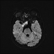
No abnormal restricted diffusion in brain parenchyma on DWI/ADC map to suggest acute infarct.

Annotated images showing the specific imaging findings in non-contrast CT brain, MRI brain: T1W, T2W, T1 post contrast and MR angiogram.
Case Discussion
Bilateral cavernous segments internal carotid arteries aneurysms are very rare occurence, where MR imaging features suggest the right sided aneurysm to be either thrombosed or ruptured with sealed intercavernous subacute hematoma as well as patent left cavernous ICA aneurysm. The mass effect of the right sided thrombosed aneurysm or intercavernous hematoma has resulted in right cranial nerve III, IV, V1, V2 and VI palsies and optic chiasm compression.
Thrombosed right internal carotid artery (C2 till C6 segment) where the right C7 segment is solely supplied by left ACA via the patent anterior communicating artery. No MR evidence of acute brain infarction.
This case shows the importance of considering aneurysm as differential diagnosis for sella turcica lesion, as internal carotid arteries pass through the cavernous sinuses.
T1-weighted, T2-weighted and post contrast MRI sequences are important sequences to look for any flow voids as black signal intensity. There are only three things black on MRI : air, bone and rapid blood flow.


 Unable to process the form. Check for errors and try again.