
Presentation
Clicking sound and locking both knees.
Patient Data
Age: 30 years
Gender: Male
From the case:
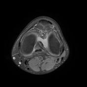
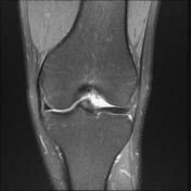
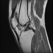
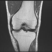
Bilateral discoid mensicus

Download
Info

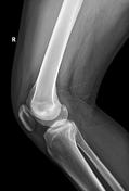
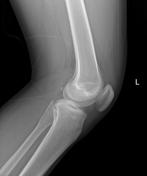
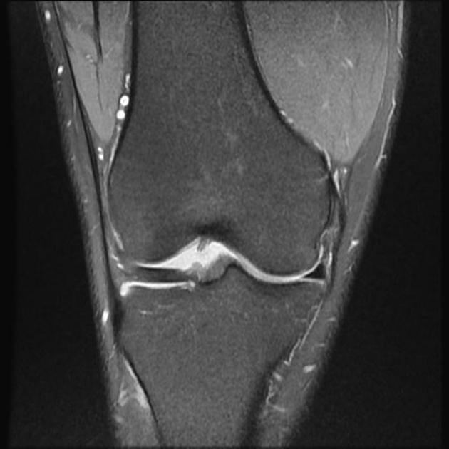
Widening of the lateral joint space is seen bilaterally and cupping of lateral tibial plateau. Additionally hypoplasia of the lateral tibial spine especially on the left knee is seen.

Download
Info
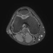
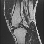
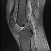
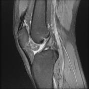
The lateral meniscus has an abnormally wide body which is seen in more than 3 sagittal images.
Download
Info

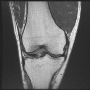
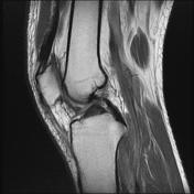
The lateral meniscus has an abnormally wide body which is seen in more than 3 sagittal images.


 Unable to process the form. Check for errors and try again.