
Presentation
Bilateral knee pain.
Patient Data
Age: 40 years
Gender: Female


Show annotations
Download
Info
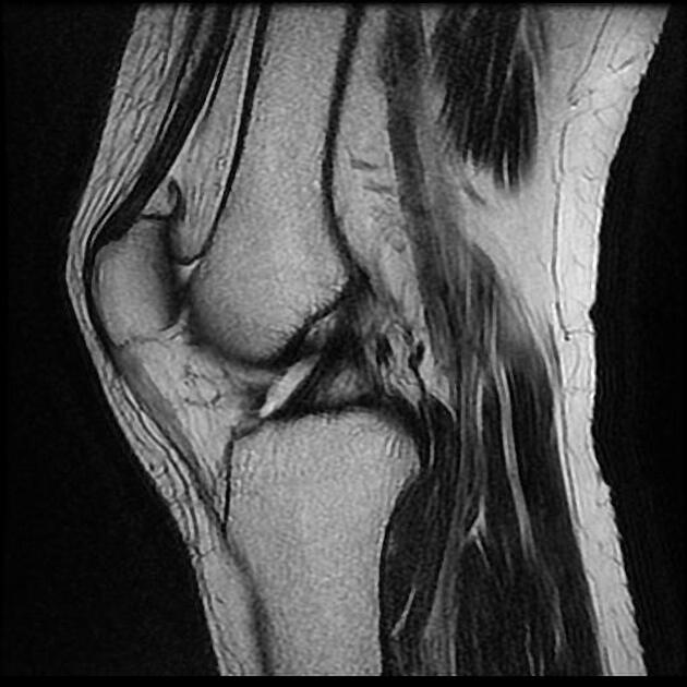
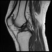
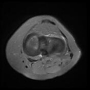
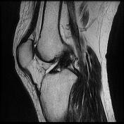
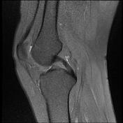
The lateral meniscus has an abnormally wide body which is seen in more than 3 sagittal images.
Show annotations
Download
Info

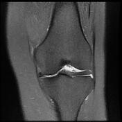
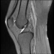
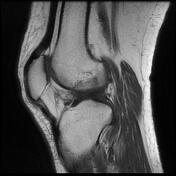
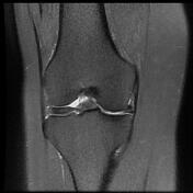
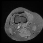
The lateral meniscus has an abnormally wide body which is seen in more than 3 sagittal images.
Case Discussion
Discoid menisci are those that have a body that is too wide, usually affecting the lateral meniscus. They are incidentally found in 3-5% of knee MRI examinations.


 Unable to process the form. Check for errors and try again.