
Presentation
Soccer player with bilateral hip pain which is becoming increasingly severe on the left.
Patient Data

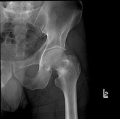
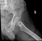
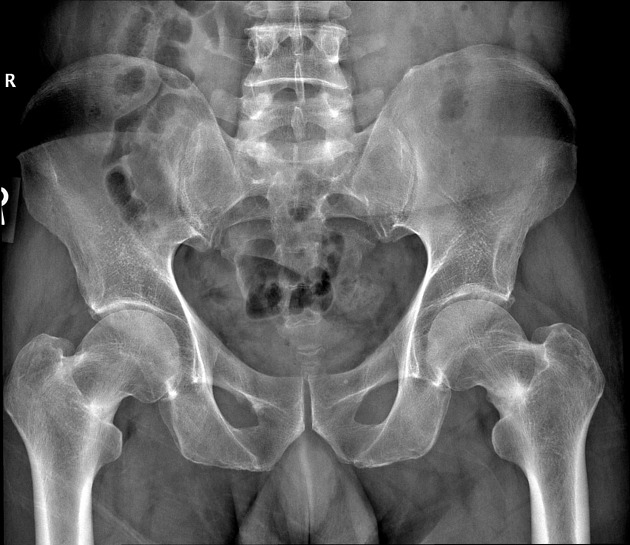
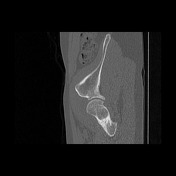
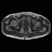
Bilaterally there is increased cortical thickness of the medial femoral necks with a linear area of central lucency, worse on the left. The left femoral neck also demonstrates cortical irregularity which is most offset at the intersection with the lucent line.
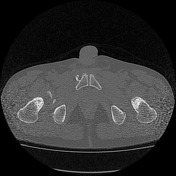

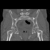
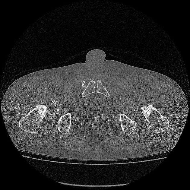
Thickening of the medial cortex of the femoral necks bilaterally which contain a linear central lucency, worse on the left.

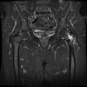
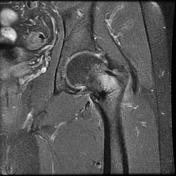
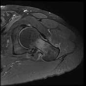
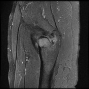
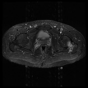

Bilaterally there are low T1/STIR-weighted signal lines within the medial portions of the femoral necks, with surrounding hyperintense STIR signal within the left femoral neck. There is associated mild surrounding soft-tissue edema on the left as well, but no evidence of retracted tendon tear.
Bilaterally there is circumferential moderate osteophyte formation of the acetabuli and femoral head-neck junctions with complex multifocal degenerative masseration of the right and left labrum.
Case Discussion
Non-displaced acute stress fracture of the left femoral neck, and chronic stress fracture of the right femoral neck.
Remember that stress fractures are caused by abnormal stress on normal bone, whereas insufficiency fractures are due to normal stress on the abnormal bone.


 Unable to process the form. Check for errors and try again.