
Bilateral middle cerebellar peduncles Wallerian degeneration secondary to unilateral pontine infarction
Presentation
Follow up of recent pontine infarction 3 months ago.
Patient Data


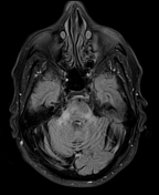

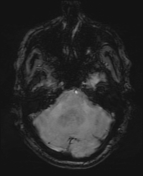
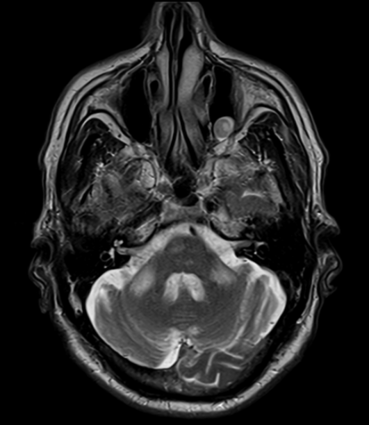
Right paramedian old pontine infarction. Hyperintensity in the middle cerebellar peduncles (MCPs).
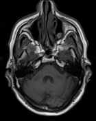
Hyperintensity in the medullary pyramid which represents corticospinal tract Wallerian degeneration.
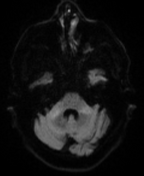
Bilateral cerebral white matter chronic ischaemic foci.
Case Discussion
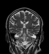
On the first MRI of the brain acquired 3 months earlier (not available), there was a recent right pontine acute infarct but with normal appearance of the cerebellum, cerebellar peduncles, midbrain, and medulla. On the current MRI, there is ageing of the right paramedian pontine infarction, with newly appreciated hyperintensities in the bilateral middle cerebellar peduncles (MCP) and the right medullary pyramid, concomitant with bilateral middle cerebellar peduncles and corticospinal tract Wallerian degeneration.
The MCP is vulnerable to Wallerian degeneration because it is the largest and main pathway for the pontocerebellar tracts. MCP degeneration appears as bilateral and symmetrical hyperintensities along the MCPs on T2-weighted and FLAIR images, usually detectable 2–3 months after stroke.
Case courtesy of Prof.Ayda.Yousif. MD of radiodiagnosis. National cancer institute. Egypt.


 Unable to process the form. Check for errors and try again.