
Patient Data
Age: 65 years
Gender: Female


Download
Info
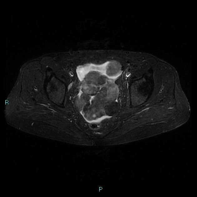
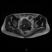
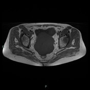
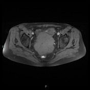
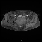
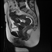
In the pelvic cavity are visible some solid formations, of elongated morphology, partly confluent, which show low signal intensity on T1 and T2 sequences, characterized by the absence of enhancement.
These lesions are separable from the uterus which is of normal-sized and shape. Is associated with peritoneal effusion of discrete entities.
Not visible ovaries.
No adenopathy.
Case Discussion
Bilateral salpingo-oophorectomy.
Histology: Bilateral ovarian fibroma.
Case courtesy: Dr.ssa Barbara Severini


 Unable to process the form. Check for errors and try again.