
Presentation
History of yellow eyes with abdominal tenderness. No previous radiological imaging as having been residing in the countryside. No history of trauma.
Patient Data
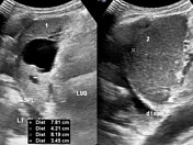
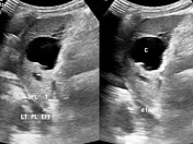
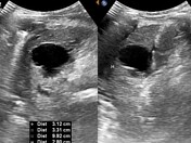
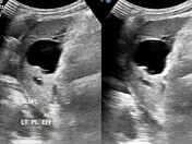
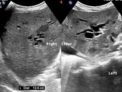
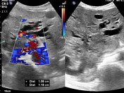
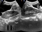

Incidentally chanced bilobar splenic organ (polysplenia) noticed during an abdominal ultrasound examination. The two splenic parenchymal plates are centrally separated evenly with a fissural linear echogenic structure cranio-caudally.
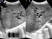
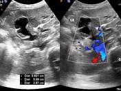
The laterally placed splenic organ (labeled 2, measuring 8.19 x 3.45 cm) shows smooth parenchymal echotexture while it's second counterpart (medially placed; labeled 1, measuring 7.81 x 4.21 cm) demonstrates an eccentric multilocular, anechoic and posteriorly enhancing thin walled cystic lesion.
Case Discussion
Bilobate (aka bipartite or bilobed) splenic organs are quite rare and are mostly congenital and mostly asymptomatic. In this illustration, the patient had no previous history of radiological imaging or trauma and presented for other unrelated clinical symptoms of which targeted abdominal ultrasound yielded:
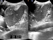
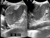
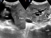
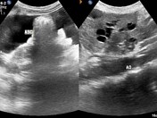
- Biliary duct ectasia with mildly thick walled gallbladder outline with intra-luminal tumefactive sludge minimally adherent within it's posteromedial lumen
- Small multiple scattered intra-hepatic hypoechoic lesions suggesting infarcts or hepatic micro-abscesses
- Small sized simple hepatic parenchymal cysts.
- Minimal ascites
- Moderate left sided pleural effusion with left basal lung consolidation and peripherally distributed miniature basal lobe air-bronchograms ipsilaterally.


 Unable to process the form. Check for errors and try again.