
Presentation
No history of trauma.
Patient Data
Age: 40 years
Gender: Female
From the case:
Bipartite scaphoid
Download
Info
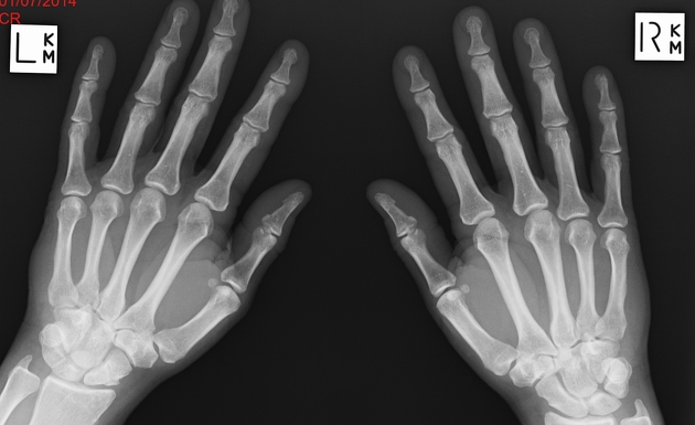
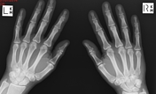
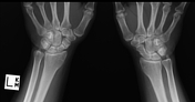
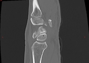
The scaphoid has ossified as two separate segments, each with well defined smooth borders. Alignment of these two segments does appear within normal limits.
From the case:
Bipartite scaphoid
Download
Info

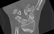
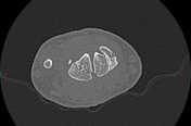
Two well defined segments of scaphoid with well corticated margins are confirmed with CT.
Case Discussion
Failure of fusion of scaphoid ossification may result in a congenital bipartite scaphoid.
Absence of trauma, equal density and size of both segments and a smooth, rounded well-defined cortex is helpful for diagnosis.


 Unable to process the form. Check for errors and try again.