
Presentation
Hematuria.
Patient Data
Age: 75 years
Gender: Male
From the case:
Bladder calculi


Download
Info
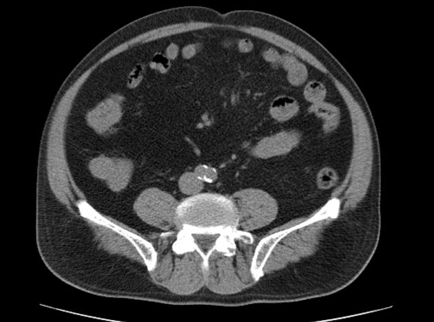
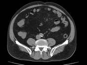
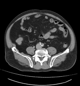
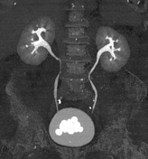
Numerous calculi of various size (from 8 to 23 mm) are noted within the urinary bladder with a mean density at 560 HU.
Normal appearance of both kidneys with no renal or ureteric stone seen.
Enlarged prostate (weight = 55 g).
Case Discussion
CT features of bladder calculi.
Bladder calculi occur either from migrated renal calculi or urinary stasis. Bladder calculi can be divided into primary and secondary stones:
- Primary: stones formed in sterile urine, usually renal calculi which have migrated down into the bladder
- Secondary: stones form de novo in the bladder or from concretions on foreign material (e.g. urinary catheters)


 Unable to process the form. Check for errors and try again.