
Presentation
Giddiness and one episode of loss of consciousness. Suspected transient ischemic attack.
Patient Data


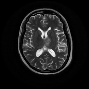
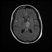
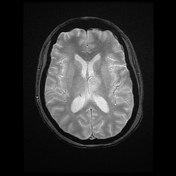

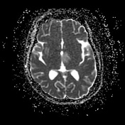

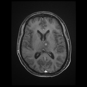
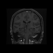
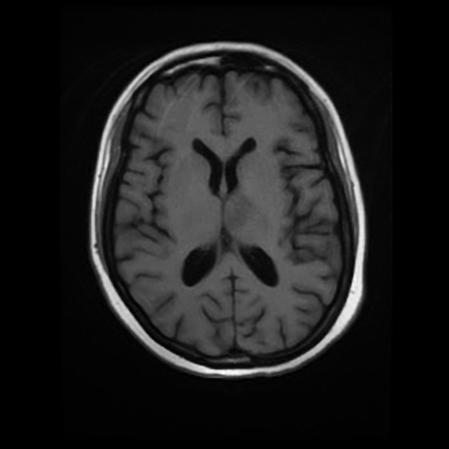
Well defined T1 hypointense, T2 hyperintense, intensely enhancing intra-axial lesions with perilesional vasogenic edema are seen in grey white matter junction of left frontal and parietal lobes, left thalamus, right paramedian dorsal midbrain and pons, right external capsule and superior temporal gyrus, left half of cerebellar vermis and right cerebellar hemisphere.
No vascular thrombosis or features of raised intracranial pressure. No acute infarction or acute intracranial hemorrhage.
Case Discussion
Brain secondaries are becoming more common thanks to better survival rates of cancer patients. Approximately 10-16 % of patients metastasize to brain, around 50 % of HER2 positive breast cancers.


 Unable to process the form. Check for errors and try again.