
Presentation
Headaches with altered level of consciousness in a patient treated for breast carcinoma.
Patient Data
Age: 40 years
Gender: Female
From the case:
Brain metastases from breast carcinoma


Download
Info

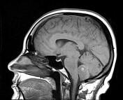
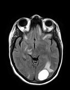
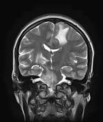
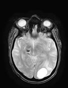
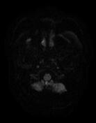
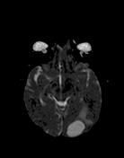
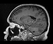
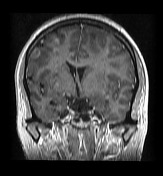
Innumerable scattered enhancing intra-axial lesions of various sizes, disseminated in both cerebellar and cerebral hemispheres and brainstem with surrounding vasogenic oedema. Some of them show intrinsic high signal on T1 and low signal on GE sequences (haemorrhagic). The largest lesion is cystic, located in the left occipital region with peripheral enhancement with no restricted diffusion. Mild midline shift to right side.
Case Discussion
MRI features of innumerable enhancing intra-axial lesions with surrounding vasogenic oedema in a patient treated for breast carcinoma, most consistent with brain metastases.


 Unable to process the form. Check for errors and try again.