
Presentation
Long-standing indolent left lateral neck mass.
Patient Data
Age: 45 years
Gender: Female
From the case:
Branchial cleft cyst


Download
Info
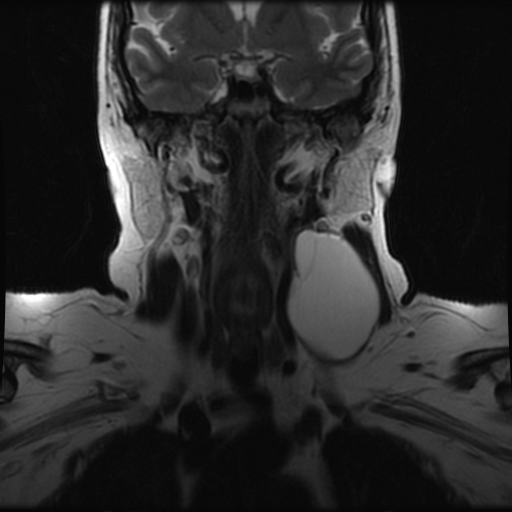
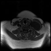
A well-defined cystic lesion in the left side of the neck deep to the left sternomastoid muscle with clear fluid content and a thin enhancing wall. It is lateral to the carotid sheath and posterior to the left submandibular gland.
Case Discussion
Imaging findings of a neck cystic lesion with the classic location for a second branchial cleft cyst. No signs of infection or complications identified.


 Unable to process the form. Check for errors and try again.