
Presentation
Trauma
Patient Data


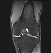
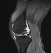
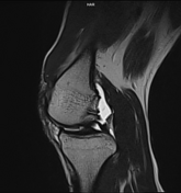
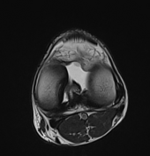
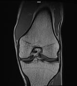
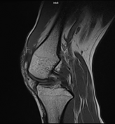
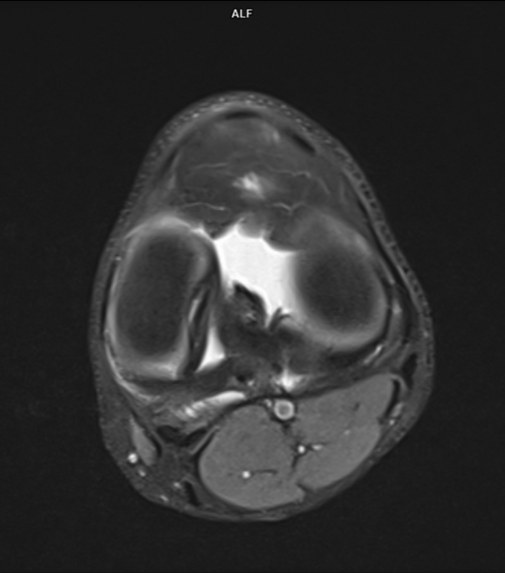
There is vertical tear of medial meniscus with medial tear segment displaced medially and is seen in the intercondylar notch in front of the posterior cruciate ligament (double PCL sign).
Mild joint effusion is seen.
ACL and PCL show normal MR morphology.
Lateral meniscus is normal in MR morphology.
Medial and lateral collateral ligaments are normal.
Distal femur, proximal tibia and fibula are normal in MR morphology. Cortical margins are preserved.
Patello-femoral and tibio-femoral joint spaces are maintained.
Patellar ligament and quadriceps tendon are normally visualized.
Popliteal vessels show normal MR morphology.
Impression: Findings are suggestive of bucket handle tear of medial meniscus.
Case Discussion
Bucket-handle meniscal tear is a type of displaced vertical meniscal tear where the inner part is displaced medially. It more commonly occurs in the medial meniscus and is often associated with anterior cruciate ligament (ACL) tear, however in this case ACL appears normal.


 Unable to process the form. Check for errors and try again.