
Presentation
The patient presented to the emergency department with three days history of right testicular pain and swelling.
Patient Data

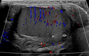
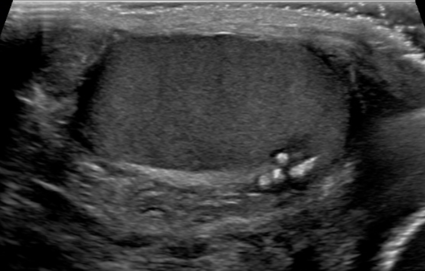
Grayscale ultrasound image of the right testis shows a cluster of coarse calcifications and a small hypoechoic area within the region of the mediastinum testis. No abnormal vascularity is identified on color Doppler imaging.


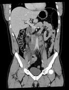
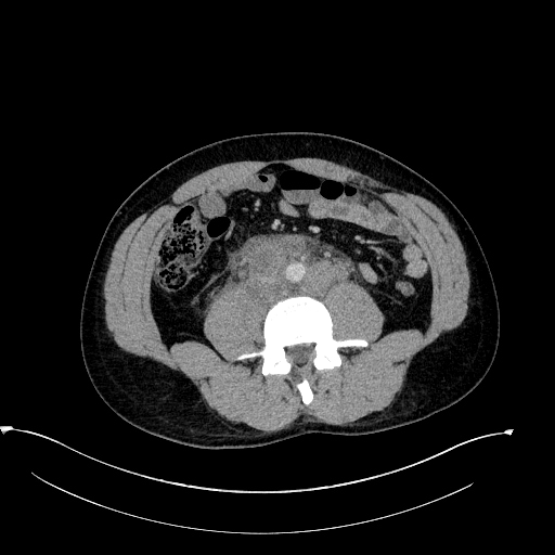
There is bulky retroperitoneal lymphadenopathy including a few necrotic lymph nodes, highly suspicious for nodal metastases.
Case Discussion
Necrotic retroperitoneal lymphadenopathy with intratesticular calcifications within a small hypoechoic area are highly suspicious of a burned-out testicular tumor in this young patient.
Biopsy of the retroperitoneal lymph nodes confirmed the diagnosis of metastatic germ cell tumor, primary appearing as embryonal carcinoma with necrosis.
A cluster of calcifications within the testis can also potentially represent phleboliths, spermatic granulomas, regions of vascular calcification, sequela of prior trauma or prior necrosis.


 Unable to process the form. Check for errors and try again.