
Presentation
Left-sided 2-3 cm mildly tender poorly mobile neck swelling.
Patient Data
Age: 40 years
Gender: Female
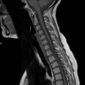
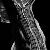
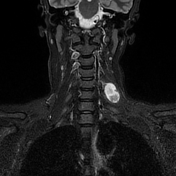
From the case:
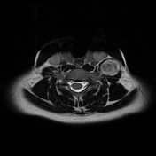
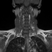
C5 nerve sheath tumour
Download
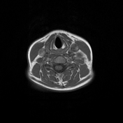
Info

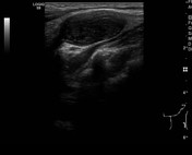
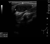
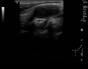
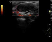
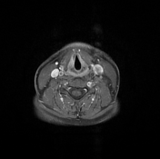
Well devoid ovoid low attenuation mass in the left side of the neck.
From the case:
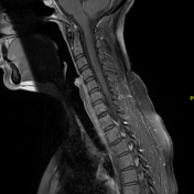
C5 nerve sheath tumour

Download
Info

3.5 cm left C5 benign appearing nerve sheath tumour. This has increased from 3 cm on the prior study.
No other abnormality.
Case Discussion
C5 nerve sheath tumour. This is most likely a schwannoma or neurofibroma. Radiologically it is often not possible to distinguish the subtype of peripheral nerve sheath tumour.
This changed only minimally over 3 years of interval imaging.
When small, they may be mistaken for lymph nodes on ultrasound.


 Unable to process the form. Check for errors and try again.