
Presentation
A female patient with left iliac fossa pain. Previous Cesarean section 5 years ago.
Patient Data


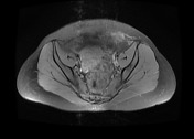
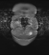
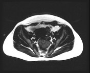
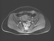
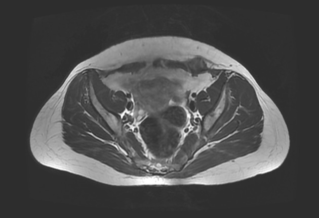
Fairly defined ovoid soft tissue mass lesion is seen in the lateral aspect of left rectus sheath being deep within subcutaneous tissue. It measures 2.2 x 2 cm. It exhibits isointense pattern on T1, hyperintense on T2 and shows inhomogeneous enhancement. It showed no fat content. The overlying subcutaneous fat is spared, no stranding and no extension beyond the under surface of the rectus sheath and peritoneal reflection.
Case Discussion
Left lower quadrant anterior abdominal wall deep subcutaneous/muscular soft tissue mass. In view of previous Cesarean section scar, patient clinical findings and current and MRI features the possibility of Cesarean scar endometriosis should be considered.


 Unable to process the form. Check for errors and try again.