Presentation
Acute, non-traumatic left shoulder pain
Patient Data
Age: 50 years
Gender: Female
From the case:
Calcific bursitis of the shoulder
![](https://prod-images-static.radiopaedia.org/images/1178724/91bc1a7be2138a0c72a19740adc72d_thumb.jpg)
![](https://prod-images-static.radiopaedia.org/images/1178731/3549b2e568734c2303e08f1f7adad6_thumb.jpg)
Download
Info
![](https://prod-images-static.radiopaedia.org/images/1178724/91bc1a7be2138a0c72a19740adc72d_big_gallery.jpg)
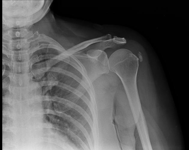
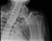
Radiographs ( AP and True AP standing ) show Periarticular calcification.
From the case:
Calcific bursitis of the shoulder
![](https://prod-images-static.radiopaedia.org/images/1178738/5b683e0fcf4b501f216226c31f8337_thumb.jpg)
![](https://prod-images-static.radiopaedia.org/images/1178745/9e5d069e8cfd31b78b8296bd4dad66_thumb.jpg)
![](https://prod-images-static.radiopaedia.org/images/1178752/13f4a793ef20a266f830b542c488ae_thumb.jpg)
![](https://prod-images-static.radiopaedia.org/images/1178759/119dbc740069e7b341633bf290dde0_thumb.jpg)
![](https://prod-images-static.radiopaedia.org/images/1178766/21660452a450b5876aeaa8df0bd534_thumb.jpg)
Download
Info
![](https://prod-images-static.radiopaedia.org/images/1178738/5b683e0fcf4b501f216226c31f8337_big_gallery.jpg)
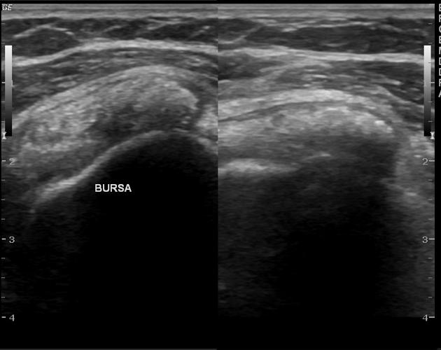
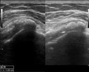
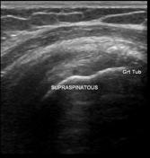
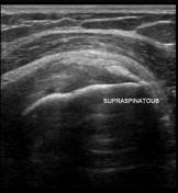
Most of calcification is in bursa with hypervascularity. Small amount of calcification in supraspinatus. Rupture of supraspinatus calcification into bursa leads to bursitis and acute pain.
Case Discussion
The findings are compatible with calcific bursitis of the shoulder.