
Presentation
Case presented with mild trauma to the back of his head. Reports history of mild dysphagia
Patient Data
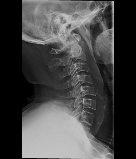
Thickening of the soft tissue planes anterior to the C1- C5 cervical vertebra.
Faint round calcification is seen below the anterior arch of C1.


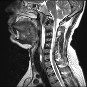
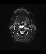
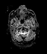
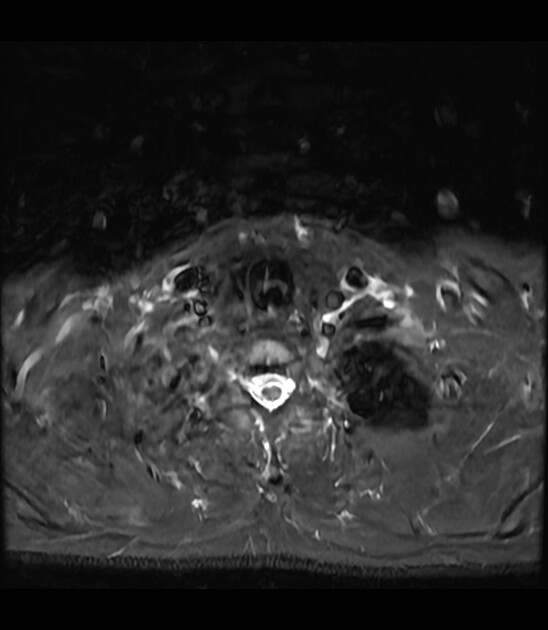
An extensive area of T2 hyperintensity is identified in the retropharyngeal space. It starts from the level of the oropharynx and extends up to the level of the epiglottis where it appears to extend laterally into the carotid spaces bilaterally. Mild mass-effect is identified.
No clear evidence of a well-circumscribed mass can be identified. No evidence of true diffusion restriction on the DWI and ADC map sequences. The findings are suggestive of retropharyngeal edema.
Calcifications at the insertion of the longus colli muscle tendons at the C1-2.
No evidence of a retropharyngeal abscess could be identified.

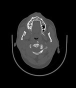

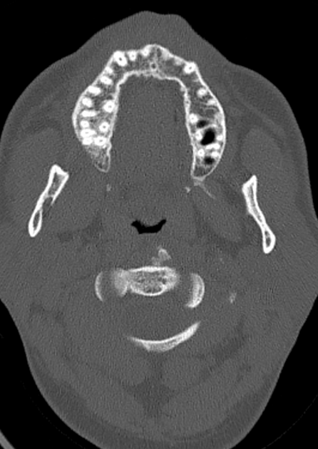
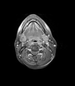
Coarse calcifications are identified at the insertion of the longus colli muscles at the C1-2 level.
Case Discussion
Calcific tendinitis of the longus colli is an inflammatory condition most commonly related to calcium hydroxyapatite deposition disease. It is a self-limiting condition that usually resolves after 2-3 weeks. Symptoms are usually treated with non-steroidal anti-inflammatory drugs.
MRI and CT images are courtesy of Dr. Georgios Kapsas.


 Unable to process the form. Check for errors and try again.