
Presentation
Stiff neck, rule out retropharyngeal abscess.
Patient Data
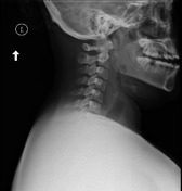
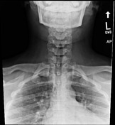

Lateral radiograph demonstrates retropharyngeal soft tissue prominence extending from C2 to C6 vertebrae measuring up to 1.8 cm at the level of C6. Lucency is also visualized in the prevertebral soft tissue at the level of C4 to C6. Findings are suspicious for retropharyngeal cellulitis/abscess. The osseous structures appear intact. No radiopaque foreign body is seen.
Not originally appreciated on this study were ossific densities projecting anterior to and at the level of the C1/odontoid process.


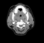

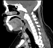

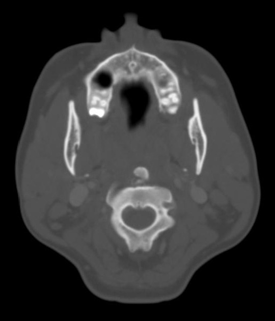
There is extensive calcification of the longus coli muscle anteriorly with evidence of fluid collection in the retropharyngeal space which measures approximately 3 cm on transverse, 1 cm on the AP and it is extending from C2 down to upper border of the C4. No evidence of soft tissue enhancement which would suggest retropharyngeal abscess. The fluid collection is related to calcific tendinitis of the longus colli muscle.
Case Discussion
The lateral radiograph demonstrated prevertebral swelling, suspicious for retropharyngeal cellulitis/abscess. Furthermore, there were ossific densities projecting anterior to and at the level of the odontoid process. This was not originally described, but this finding provides an additional differential diagnosis of calcific tendinitis of the longus colli muscle. Other differentials include retropharyngeal hematoma in the correct clinical setting or mass such as hemangioma.
The CT clarified that the retropharyngeal fluid collection was secondary to calcific tendinitis of the longus colli muscle.


 Unable to process the form. Check for errors and try again.