
Presentation
Acute right hip pain.
Patient Data
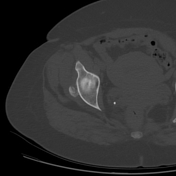

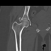
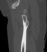
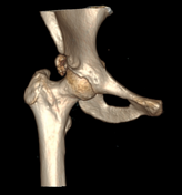

Irregular calcifications enrolling the right acetbular roof (at the anatomical location of the reflected head of the rectus femoris muscle).
Case Discussion
Acute calcific tendinitis of the rectus femoris muscle is a rare entity. It is caused by an acute inflammatory reaction secondary to calcium hydroxyapatite crystal deposition. The shoulder is mostly involved, less commonly the wrist, elbow, and knee, and rarely the longus colli and hip joint can also be affected. Calcifications around the hip joint might be at the greater trochanter, gluteus medius/minimus insertion, and the rectus femoris muscle. Calcifications appear on plain X-ray and CT scans and might disappear on follow-up radiological examination. On MRI, calcifications appear of dark signal on all pulse sequences wth surrounding soft tissue and marrow edema.


 Unable to process the form. Check for errors and try again.