
Presentation
Non-specific right hypochondrial pain
Patient Data
Age: 65 years
Gender: Female
From the case:
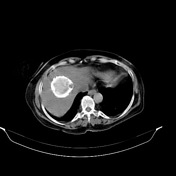
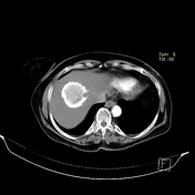
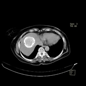
Calcified hydatid cyst


Download
Info

Four phases study of the abdomen revealed a heavily calcified rounded lesion at the right hepatic lobe which shows no evidence of enhancement or washout in different phases. The liver itself shows fatty changes yet with regular contour and homogeneous density and no evidence of cirrhotic changes. Small right renal stone and left renal cyst are also noted.
Case Discussion
Benign-looking peripherally calcified cystic lesion within the liver (or spleen) is most in keeping with a hydatid cyst. Note the small dots of calcification at the periphery of the lesion which corresponds to scolices (daughter cysts).


 Unable to process the form. Check for errors and try again.