
Presentation
Day 1 post op knee replacement for advanced osteoarthritis.
Patient Data

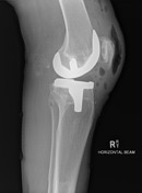
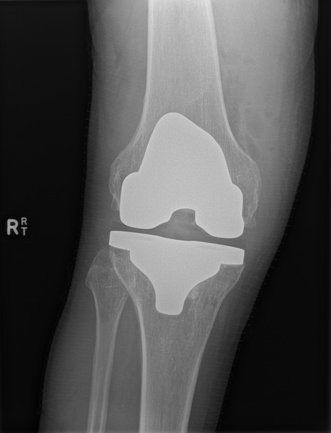
Knee joint effusion and gas and subcutaneous gas in keeping with recent total knee replacement. The femoral and tibial components are well aligned. Patellar resurfacing noted.
Large well defined corticated densities posterior to the knee joint on the lateral projection - DDX of calcified muscular hematoma, calcified loose bodies in a Baker cyst or calcified lymph nodes.

Advanced osteoarthritis. Moderate joint effusion. Fluid surrounding the popliteus tendon.
Large complex Baker cyst, with calcificed loose bodies within it.
Case Discussion
Calcified loose bodies can uncommonly be found in a Baker cyst, either arising from the knee joint and moving into the cyst (with causes including trauma, destructive arthropathy or synovial osteochondromatosis) or may arise within the cyst itself by chondrometaplasia.


 Unable to process the form. Check for errors and try again.