
Presentation
Left forehead lump. Present for two years.
Patient Data
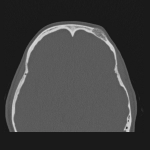
Expansile lucent lesion of left frontal bone with 'sunburst' trabecular thickening with extension beyond the external plate of the calvarium - concerning for 'hair-on-end' periosteal reaction. No other bony lesion.
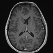
Soft tissue window of the brain (not shown) is unremarkable.
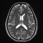

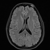
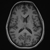

The left frontal bone lesion has heterogenous signal characteristics but is largely:
T2: hyperintense
T2: FLAIR hyperintense
T1: hypointense with speckled foci of signal hyperintensity (likely to represent fat)
T1 C+: avid contrast enhancement following gadolinium
Unremarkable brain and CSF spaces. No other lesion.
Pathology
Bone with large dilated vascular spaces within the marrow spaces. The appearances are of a cavernous hemangioma of bone.
Case Discussion
Intraosseous hemangiomas are benign vascular lesions. They have a relatively indolent clinical course however can present with lumps. Imaging appearances can be concerning, particularly the pseudo 'hair-on-end' appearance. This lesion underwent open biopsy and pathology confirmed benign hemangioma.


 Unable to process the form. Check for errors and try again.