
Presentation
Microscopic hematuria. No flank pain, nausea, vomiting or fever.
Patient Data


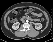
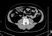
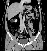
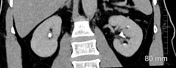
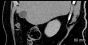
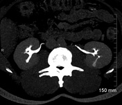
Kidneys are normal in site, size and parenchymal density. No radiopaque renal/ureteric calculi or hydroureteronephrosis is seen. Kidneys show homogeneous parenchymal enhancement on the postcontrast study.
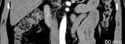
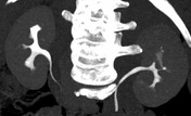
Type 1 retroaortic left renal vein (anatomical variant); otherwise, patent and well-opacified renal arteries and veins.
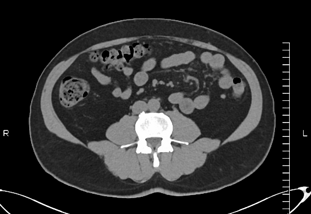
A simple cortical cyst measuring 8 x 9 mm is noted in the right kidney. No suspicious renal mass lesion is noted.
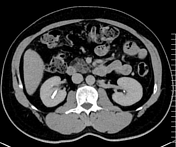
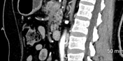
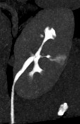
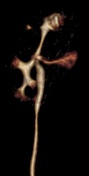
A small linear hypodensity is seen along the posterior mid-pole of the left kidney on the nephrographic phase, which shows filling with contrast in the excretory phase. No contrast leakage/extravasation is noted. No perinephric fat stranding or collection is seen.
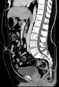
Small fat-containing umbilical hernia.
Aberrant replaced right hepatic artery (anatomical variant) arising from the SMA and coursing between the portal vein and IVC.
Case Discussion
CT scan features consistent with a small left renal calyceal diverticulum. Its main differential diagnosis is renal cyst. Both lesions (calyceal diverticulum and renal cyst) appear hypodense on the nephrographic phase of the study and cannot be differentiated from each other; however, this differentiation can be easily made on the renal excretory phase of the study when the calyceal diverticulum shows filling with contrast due to its communication with the collecting system whereas cyst does not show any filling due to lack of any connection with the collecting system.


 Unable to process the form. Check for errors and try again.