
Presentation
Recurrent left renal angle pain
Patient Data
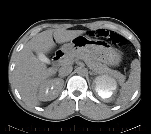
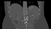
Large left renal cystic lesion with dependent calculus. Progressive contrast fill-in with time - corticomedullary phase, 10 min excretory, 60 min, 24 hours. The last image is a non-contrast CT one year later - and it shows that an extra calculus has developed. Findings in keeping with calyceal diverticulum and stasis calculi.

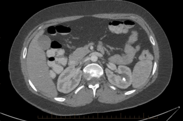
Small left renal cystic lesion with posterior calcifications. It was initially diagnosed as Bosniak Class 2 cyst. Careful windowing shows that those calcifications are likely dependent calculi instead. This would suggest this lesion being a calyceal diverticulum.
Case Discussion
Learning points:
1. Very delayed excretory phase (1-24hr) will help confirm diagnosis if in doubt.
2. Careful windowing to examine the dependent calcifications will help show that those are calculi rather than mural.


 Unable to process the form. Check for errors and try again.