
Presentation
Five months history of abdominal pain, fatigue and weight loss. The ultrasound exam revealed numerous hepatic nodules. An MRI exam was requested for characterisation.
Patient Data


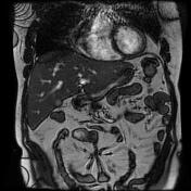
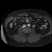
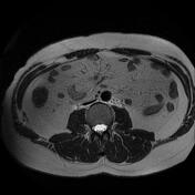
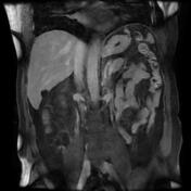
Heterogeneously enhancing soft tissue mass within the small bowel mesentery in the mid-abdomen encasing branches of the SMA with a “spoke-like” appearance of mesenteric vessels and mild distortion of the adjacent small bowel loops. A small enhancing nodule of the adjacent bowel loop is noted (best seen on the delayed arterial phase).
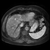
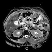
Multiple hypervascular hepatic nodules of various sizes with restricted diffusion, the largest is located in the left lobe.
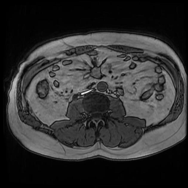
A small simple hepatic cyst is noted in segment VI.
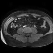
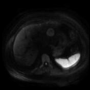
Right hydroureteronephrosis due to ureteral stone (confirmed by ultrasound).
Case Discussion
The CT features are most consistent with small bowel carcinoid tumour with mesenteric and hepatic metastases.
Small bowel carcinoid tumours, also called small bowel neuroendocrine tumours (SBNETs) are considered the most common gastrointestinal neuroendocrine tumours and most frequently involve the terminal ileum.


 Unable to process the form. Check for errors and try again.