
Presentation
Right ventricular mass noted incidentally on echocardiography. No significant past medical history.
Patient Data
Age: 65 years
Gender: Female

Download
Info

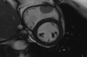
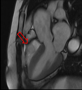
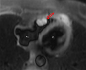
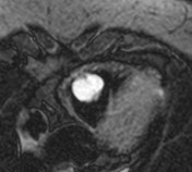
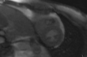
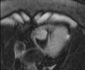
MRI images of a biopsy-proven cardiac hemangioma.
Intraluminal mass arising from the RV free wall, hyperintense in T2 weighted images with classic hemangioma-like discontinuous nodular enhancement.
Case Discussion
Cardiac hemangioma is an uncommon benign cardiac tumor (5-10%). They have no typical age of presentation and no typical cardiac chamber predilection.
The appearance, in this case, is typical with hyperintense T2 signal and gradual postcontrast enhancement on the perfusion sequence.


 Unable to process the form. Check for errors and try again.