
Presentation
Pulsatile left exophthalmos with subconjunctival haemorrhage.
Patient Data
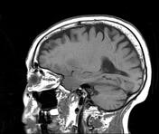

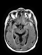
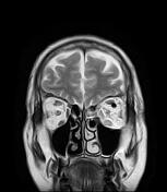
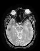
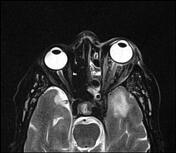
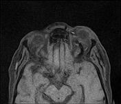
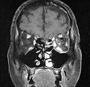


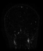
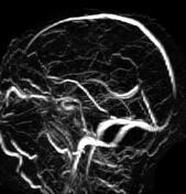

The MRI sequences demonstrate:
-
left orbital congestion
axial proptosis, grade 2
retrobulbar oedema with fat stranding
swelling of extraocular muscles
-
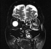
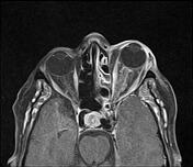
venous engorgement and enhancement
enlarged left superior ophthalmic vein with a signal void indicating a high-velocity flow (arterialised flow)
asymmetric enhancement with engorgement of the left cavernous sinus showing an attenuation similar to that of ICA
dural thickening and enhancement of the left cerebral hemisphere and along the tentorium cerebelli with white matter oedema
Case Discussion
MRI features of a caroticocavernous fistula (CCF).
Caroticocavernous fistulas (CCF) represent an abnormal communication between the carotid circulation and the cavernous sinus. They can be classified into two types: direct and indirect, according to their various features.


 Unable to process the form. Check for errors and try again.