
Presentation
Two day history of worsening diplopia.
Patient Data
Note: This case has been tagged as "legacy" as it no longer meets image preparation and/or other case publication guidelines.
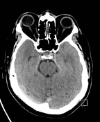
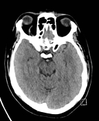
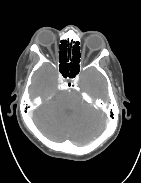
Non-contrast CT head demonstrates right proptosis and an enlarged and hyper-attenuating right superior ophthalmic vein. Differential considerations include carotid-cavernous fistula or thrombosis of the superior ophthalmic vein. The patient proceeded to a CT cerebral angiogram for further evaluation.


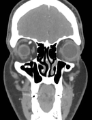
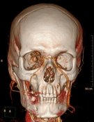
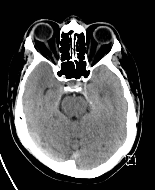
CT cerebral angiogram demonstrates early filling of the right cavernous sinus with associated enlargement of both the superior and inferior ophthalmic veins compatible with the diagnosis of carotid-cavernous fistula. The raised venous pressure is transmitted to the ipsilateral angular vein which interestingly drains through a transnasal branch to the contralateral enlarged left angular and facial veins.
Case Discussion
Carotico-cavernous fistulas represent abnormal communication between the carotid circulation and the cavernous sinus. They can be classified as direct or indirect which are separate conditions with different etiologies.


 Unable to process the form. Check for errors and try again.