
Presentation
Headache
Patient Data
Age: 40 years
Gender: Male
From the case:
Cavernoma


Download
Info
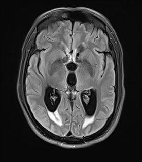
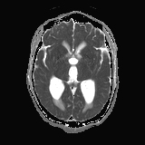
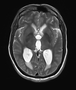
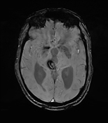
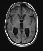
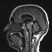
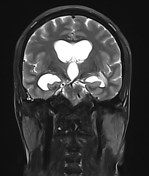
Well defined lesion containing haemorrhage in chronic and subacute stages, measuring approx 17 x 16 x 17mm is seen in posterior aspect of midbrain on right side. Mild perilesional oedema is seen. It is causing compression of aqueduct of sylvius, quadrigeminal cistern posteriorly, resulting in mild to moderate hydrocephalus and periventricular seepage.
Case Discussion
Findings are suggestive of midbrain cavernoma causing hydrocephalus.


 Unable to process the form. Check for errors and try again.