
Presentation
Chinese man presenting with weakness.
Patient Data


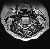
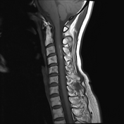
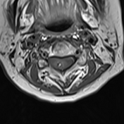
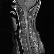
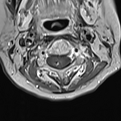
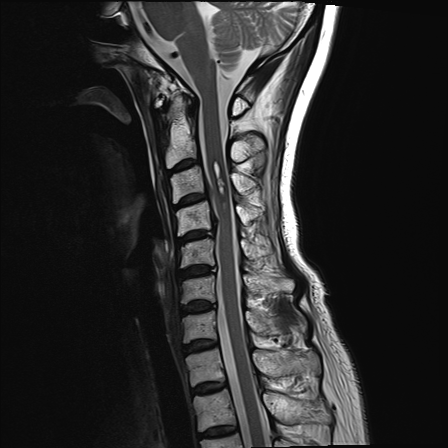
A lesion in the cord at the level of C3 is located centrally and to the right of the midline and surrounded by significant signal loss, consistent with haemosiderin. It also demonstrates intrinsic high T1 signal on non-contrast T1W images and enhancement on delayed images.
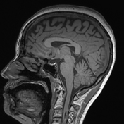
The bone marrow is uniformly fatty, more than expected for a 55-year-old. Similarly, the parotid glands are markedly atrophic, replaced by fat.
Features suggest prior irradiation.
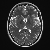
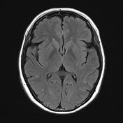
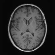
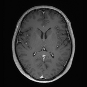
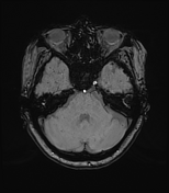
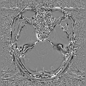
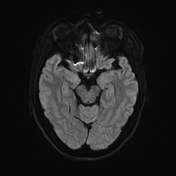
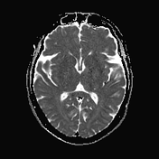
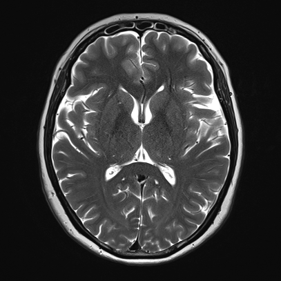
Scattered punctate foci of susceptibility artifact within the cerebellar hemispheres and throughout the temporal poles. No abnormal parenchymal diffusion restriction or contrast enhancement.
Prior bilateral maxillary antrostomies and generalised mucosal thickening.
Conclusion:
Extensive tiny punctate foci of susceptibility artifact within the cerebellar hemispheres and throughout the temporal lobes are suggestive of prior radiotherapy, presumably for nasopharyngeal carcinoma.
Case Discussion
Further history eventually obtained confirmed a history of nasopharyngeal cancer 20 years ago, treated with chemotherapy and radiotherapy.
The clues, in this case, were the distribution of microhaemorrhages, the fatty replacement and other radiation-induced changes, as well as the ethnicity of the patient.
Nasopharyngeal carcinoma is a fairly rare malignancy in most of the western world (annual incidence of less than 1 per 100,000) but has far higher prevalence, particularly among Chinese (up to 25 per 100,000), South-East Asian, and some Middle Eastern populations 1.


 Unable to process the form. Check for errors and try again.