
Presentation
Head aches
Patient Data
Age: 16 years
Gender: Male
Download
Info
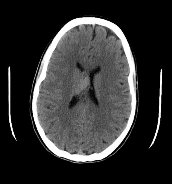
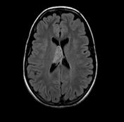
Single slice of the brain through the lateral ventricles demonstrates a slightly hyperdense mass in the body of the right lateral ventricle.


Download
Info
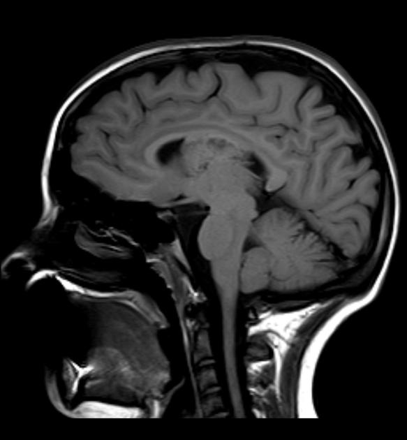
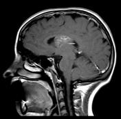
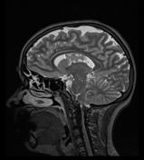
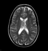
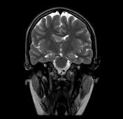
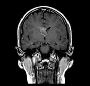
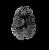
Intraventricular mass located in the body of the right lateral ventricle is heterogeneous in signal on all sequences, demonstrating only patchy enhancement.
This patient went on to have a resection which confirmed the diagnosis of a central neurocytoma, albeit with a high mitotic index.
Case Discussion
Typical appearances of a central neurocytoma.


 Unable to process the form. Check for errors and try again.