
Presentation
Headaches.
Patient Data


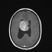
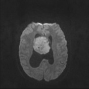
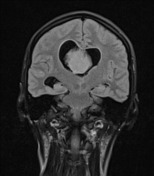
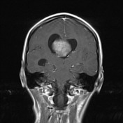
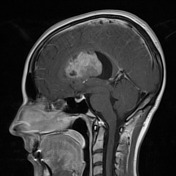
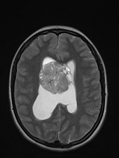
A large intraventricular mass appears to be attached to the septum pellucidum. The mass occludes the foramen of Munro with enlargement of the lateral ventricles and effacement of the overlying sulci. The mass enhances heterogeneously but relatively vividly. The mass demonstrates restricted diffusion (~500 x 10-6 mm2/s on ADC - not shown)
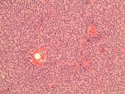
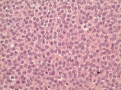
MICROSCOPIC DESCRIPTION:
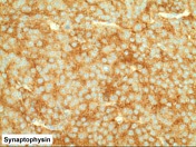
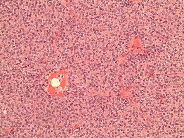
Paraffin sections show fragments of a densely hypercelullar tumor. This consists of diffuse sheets of cells with uniform round and oval nuclei with an open chromatin arrangement, conspicuous nucleoli and delicate neuropil-like processes. These show nuclear immunostaining for NeuN (not shown), cytoplasmic staining for beta-tubulin (not shown) and membrane staining for synaptophysin. No staining for GFAP is seen. Occasional mitotic figures are identified (2/20HPF). No necrosis is seen. The features are of central neurocytoma. The topoisomerase labeling index is approximately 5%
DIAGNOSIS: Central neurocytoma (WHO Grade II).
Case Discussion
The patient underwent a craniotomy and the mass was resected, confirmed pathologically to be a central neurocytoma. Only a year later the mass recurred, and again was resected and again a central neurocytoma was proven histologically.
The degree of enhancement and diffusion restriction are a little atypical and probably relate to the relatively aggressive recurrence.


 Unable to process the form. Check for errors and try again.