
Presentation
Headache.
Patient Data


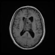
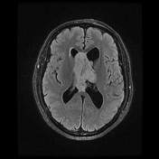
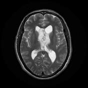
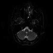
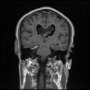
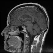
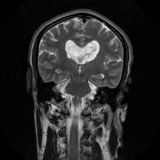
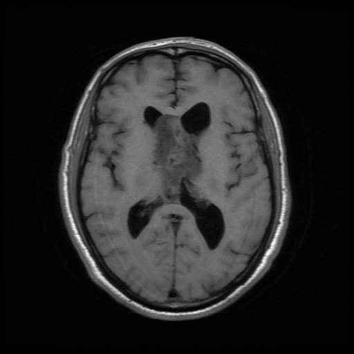
Large intraventricular mass attached to the left side of the septum pellucidum, filling and expanding the body of the both lateral ventricles, not reaching the third ventricle. It exhibits high T2 signal, low T1 signal with multiple small cystic regions (bubble appearance). Following contrast administration only minimal heterogeneous enhancement is demonstrated . There is mild dilatation of the lateral ventricles (hydrocephalus) without transependymal edema. The mass restricts on diffusion images.
Case Discussion
Central neurocytoma characteristically occurs in the lateral ventricle, extension into the third ventricle may be demonstrated. Nearly 50% of central neurocytomas are seen in the anterior part of a lateral ventricle within the foramen of Monro region, 15% in both lateral and third ventricles and 15% are bilateral.
Intraventricular oligodendroglioma or ependymoma may be identical to central neurocytoma without immunohistochemical studies. All of them appear hyperattenuating to the white matter on CT images with calcification in most of them.


 Unable to process the form. Check for errors and try again.